
Fig. 5
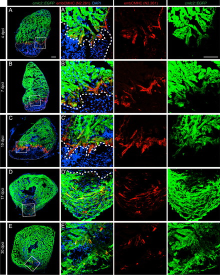
N2.261 immunostaining reveals a reactivation of embCMHC expression at the vicinity of the post-infarcted tissue. (A–E) Representative images of cmlc2::EGFP hearts at different time points during regeneration. Post-infarcted tissue is encircled by a dashed line. (A′–E′) Higher magnifications of the framed areas shown in (A-E). EmbCMHC (red) expression was detected only in the vicinity of the cryoinjury border, where CMs dedifferentiate and reduce cmlc2 expression. At 4 dpci (A), a few scattered embCMHC-positive cells emerge along the injury. At 7 (B) and 10 (C) dpci, the embCMHC-positive cells form a continuous zone above the post-infarcted tissue. At 17 dpci (D), embCMHC-positive cells intermingle with the mature CMs. At 30 dpci (E) only few embCMHC- positive cells are present within the new myocardium. Scale bar (A, A100=(′ µm. |
Reprinted from Developmental Biology, 399(1), Sallin, P., de Preux Charles, A., Duruz, V., Pfefferli, C., Jazwinska, A., A dual epimorphic and compensatory mode of heart regeneration in zebrafish, 27-40, Copyright (2015) with permission from Elsevier. Full text @ Dev. Biol.