
Fig. 1
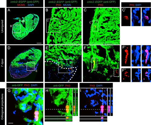
Determination of the G1/S-phase and the mitotic indexes of cardiac and non-cardiac cells in the zebrafish ventricle. Heart sections were quadruple labeled using DAPI (nuclei) and antibodies against GFP (cmlc2::EGFP, cardiac cells), MCM5 (G1/S-phase) and PH3 (mitosis). (A–C) Uninjured heart contains a few proliferating cells. (B–C) Single PH3-positive mitotic cell (red) within the intact myocardium demarcated by GFP (green). (Có) Higher magnification of the framed area in (C) with an overlap between PH3 (red) and DAPI (blue). (D–I) Heart at 7 dpci displays enhanced cell proliferation in both the post-infarction area and the remaining myocardium. (E, F) PH3-positive mitotic cells (red) at the injury border in the myocardium (green) and in the post-infarcted tissue (GFP-negative, black). (F′,F′′) Higher magnification of the framed areas in (F) with an overlap between PH3 (red) and DAPI (blue). Fragmented pattern of nuclear staining reveals mitotic segregation of condensed chromosomes. (G–I) Representative image of a mitotic cardiac cell. Orthogonal projections demonstrate a co-localization between PH3 (red), GFP (green) and DAPI (blue) staining. Scale bars in (A, B, C)=100 µm; in (G)=10 µm. |
Reprinted from Developmental Biology, 399(1), Sallin, P., de Preux Charles, A., Duruz, V., Pfefferli, C., Jazwinska, A., A dual epimorphic and compensatory mode of heart regeneration in zebrafish, 27-40, Copyright (2015) with permission from Elsevier. Full text @ Dev. Biol.