
|
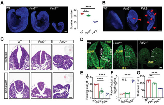
Figure 1
Pak2−/− embryos fail to develop DLHPs. A) Numbers of somite in WT, Pak2 +/−, and Pak2 −/− embryos at E9.5. (F (2, 9) = 188.2, p < 0.0001; p < 0.0001 for WT versus Pak2 −/− mice, p = 0.0034 for WT versus Pak2 +/− mice). B) Pak2 −/− embryos exhibit a characteristic phenotype of craniorachischisis, with an open neural tube (indicated by asterisks) extending from the forebrain to the posterior spinal cord. C) Hematoxylin/eosin‐stained hindbrain and spinal cord of WT and Pak2 −/− embryos at E9.5. D) Quantification of the neural tube of WT, Pak2 +/−, and Pak2 −/− embryos at E9.5. E–G)The PAK2 levels (E, F (2, 9) = 72.68, p < 0.0001; p < 0.0001 for WT versus Pak2 −/− mice, p < 0.0001 for WT versus Pak2 +/− mice), the ratio of apical to basal surface at the DLHP (F, WT, 0.547 ± 0.034; Pak2 −/− mice, 2.397 ± 0.067; Pak2 +/− mice, 0.640 ± 0.037; F (2, 9) = 460.7, p < 0.0001; p = 0.0001 for WT versus Pak2 −/− mice, p = 0.335 for WT versus Pak2 +/− mice), and the thickness of neural tube (G, p = 0.0001 for WT versus Pak2 −/− mice, p = 0.003 for WT versus Pak2 +/− mice) of WT, Pak2 +/−, and Pak2 −/− embryos at E9.5. One‐way ANOVA with Dunnett's multiple comparisons, n = 4 embryos for each genotype (A, E, F, G). Scale bar: 200 µm (A,B); 20 µm (C,D).