
|
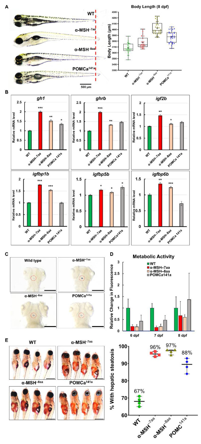
Figure 3
The level of α-MSH regulates normal somatic growth and energy balance in zebrafish embryos/larvae. (A) Left: Lateral view of wild-type and pomca mutant larvae at 8 dpf. Scale bar = 500 μm. Right: Statistical analysis of BL (jaw to tail fin) in WT controls and pomca mutant larvae at 8 dpf. Data are shown as the mean ± SD (n = 30). (B) Expression of GH/IGF axis genes, gh1, ghrb, igf2b, igfbp1b, igfbp15b, and igfbp6b, in WT controls and pomca mutants larvae at 8 dpf. Values are the mean ± SEM. * p < 0.05, ** p < 0.01, and *** p < 0.001 compared with WT groups. (C) Whole-mount ISH showing the increased expression of gh1 in the pituitary in pomca mutants larvae at 5 dpf. Scale bars = 200 μm. (D) Response to depletion of α-MSH in pomca mutant larvae by the Alamar Blue assay. Three larvae per well were incubated in 4 mM sodium bicarbonate with 1% Alamar Blue. The fluorescence of the solution was measured at different time points. Data are reported as the relative change in fluorescence intensity at least three times independently. (mean ± SEM, n = 30). (E) Left: Hepatic steatosis was observed by whole-body Oil Red O staining in pomca mutant larvae at 21dpf. Right: Percentages of WT and pomca mutant larvae with strong levels of hepatic steatosis at 21 dpf were calculated from at least 50 fish in each group. Data were representative of four independent experiments. Scale bar = 1 mm.