
|
Figure 1
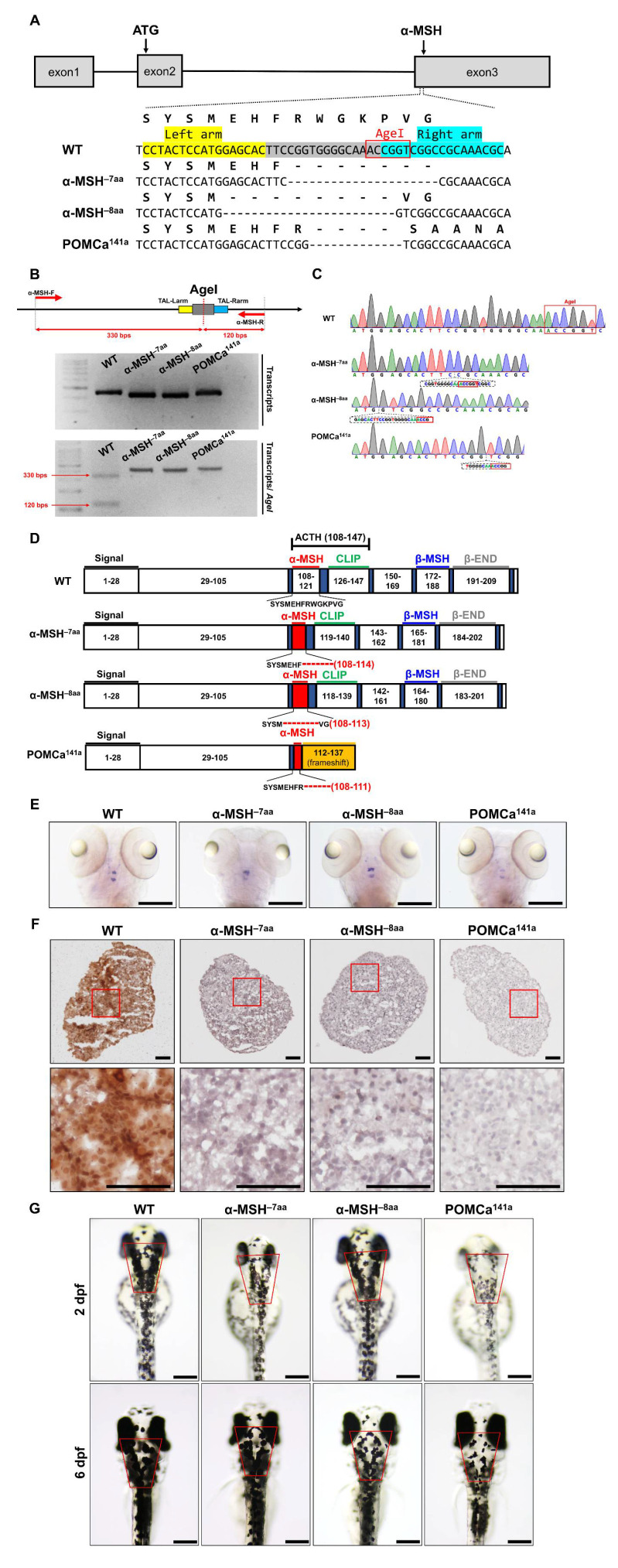
Generation of zebrafish pomca mutants using TALEN. (A) Schematics of the three pomca mutant alleles generated. The sequences in the third exon that were targeted by the TALEN pairs are shown in gray boxes. The AgeI restriction enzyme recognition site for genotyping purposes is shown in a red box; dashed lines indicate deleted nucleotides. The left and right TALEN targeting sites are highlighted in yellow and sky blue, respectively. (B) Upper: Schematic illustration of the primers used for RT–qPCR detection of mutations. The specific primer is to the mutated site/scheme of the locations of primers (red arrows) designed to detect a disruption in the spacer of the third exon. Yellow box, left TALEN sites (TAL-L); sky blue box, right TALEN sites (TAL-R); grey box, spacer; red dashed line, AgeI restriction site. Lower: Results of RT–qPCR analyses on fin clips of heterozygous F1 fish containing one of the corresponding mutations (as indicated in A). (C) Chromographs illustrating the sequences in the third exon of the pomca WT controls and the nucleotide deletion of pomca mutants. The boxed sequence in red indicates the restriction enzyme AgeI cutting sites in WT controls that were deleted in the mutant fish. (D) These diagrams show the predicated Pomca protein of pomca mutants compared with the WT Pomca protein. (E) Whole-mount ISH showing the expression of pomca transcripts in the pituitary in WT controls and pomca mutants larvae at 5 dpf. Scale bars = 200 μm. (F) Expression patterns of α-MSH in 12 mpf Pomc neuron samples after Immunohistochemistry-frozen section (IHC-F) staining. Scale bar = 50 μm. (G) Dorsal view of 2 dpf and 6 dpf pomca mutant larvae. Scale bars = 200 μm.