FIGURE

Figure 6.

ID
ZDB-FIG-200829-69
Publication
Liu et al., 2020 - TMEM216 Deletion Causes Mislocalization of Cone Opsin and Rhodopsin and Photoreceptor Degeneration in Zebrafish
Other Figures
All Figure Page
Back to All Figure Page
Figure 6.

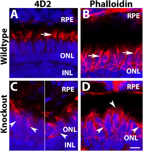
Defective organization of F-actin in tmem216 knockout photoreceptors. Cryosections were immunostained with 4D2 (red). Some sections were stained with Phalloidin-RITC for F-actin (red). (A, C) 4D2 staining of wildtype and tmem216snyΔ175 homozygous retina. 4D2 labels rod outer segments in the wildtype zebrafish (arrow in A); however, its reactivity was frequently mislocalized to the rod cell body in the knockouts (arrowheads in C). (B, D) Phalloidin staining in wildtype and tmem216snyΔ175 homozygous retina. Vertically oriented F-actin in the wildtype (arrows in B) were disrupted in tmem216snyΔ175 homozygous retina (arrowheads in D). Scale bar in H: 6 µm.

Expression Data
Antibody:
Fish:
Anatomical Terms:
Stage: Days 14-20

Expression Detail
Antibody Labeling
Phenotype Data
Fish:
Observed In:
Stage: Days 14-20

Phenotype Detail
Acknowledgments
This image is the copyrighted work of the attributed author or publisher, and ZFIN has permission only to display this image to its users. Additional permissions should be obtained from the applicable author or publisher of the image. Full text @ Invest. Ophthalmol. Vis. Sci.