
Fig. 3
- ID
- ZDB-FIG-200727-16
- Publication
- Sinagoga et al., 2020 - Mitf-family transcription factor function is required within cranial neural crest cells to promote choroid fissure closure
- Other Figures
- All Figure Page
- Back to All Figure Page
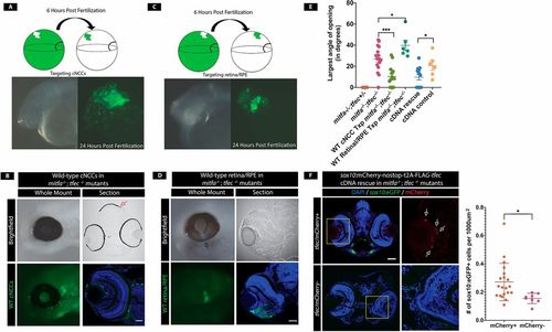
mitfa and tfec are required within cNCCs to promote CF closure. (A) NCC transplantations. At 6 hpf, wild-type dextran-labeled cells are transplanted into the cNCC-fated region of a mitfa−/−;tfec−/− embryo. At 24 hpf, transplanted cells are detected in the cNCC population. (B) Example of 4 dpf wild-type (WT) cNCC transplanted mitfa−/−;tfec−/− embryo in which the coloboma has been rescued. Transplanted cNCCs are detected in and around the eye and rescue cNCC-derived pigmented melanocytes (red arrow) confirming successful transplantation. Scale bar: 25 µm. (C) Retina/RPE transplants. At 6 hpf, wild-type dextran-labeled cells are transplanted into the RPE/retinal-fated region of a mitfa−/−;tfec−/− mutant. At 24 hpf, transplanted cells are detected within the eye, indicating successful transplantation. (D) At 4 dpf, transplanted embryos still possess colobomas. Arrow indicates a coloboma in the retina/RPE transplanted eye. (E) Quantification of colobomas (largest angle of CF opening) in rescue experiments. CF opening is significantly reduced in mitfa−/−;tfec−/− embryos transplanted with wild-type cNCCs (P=0.0004, n=14 eyes). However, colobomas are not resolved in embryos transplanted with wild-type retina/RPE cells and are actually larger than in non-transplanted control mitfa−/−;tfec−/− mutants (P=0.037, n=6 eyes). tfec cDNA injection also rescues colobomas (P=0.033, n=14 eyes). (F) pDestTol2pA2-sox10:mCherry-nostop-t2A-FLAG-tfec-pA injection reduces CF opening in mitfa−/−;tfec−/− mutants. Arrows indicate mCherry+ (rescued) neural crest cells. Quantification of sox10:eGFP+ cells shows rescue of cNCCs within the eye field (P=0.023, n=20 mCherry+, n=8 mCherry− eyes). *P<0.05, ***P<0.001. Dorsal is upwards in all images. Data are mean±s.e.m. Scale bars: 25 μm (B,D); 50 μm (F). |