
Fig. 2
- ID
- ZDB-FIG-190606-5
- Publication
- Kelu et al., 2018 - Characterization of ADP-ribosyl cyclase 1-like (ARC1-like) activity and NAADP signaling during slow muscle cell development in zebrafish embryos
- Other Figures
- All Figure Page
- Back to All Figure Page
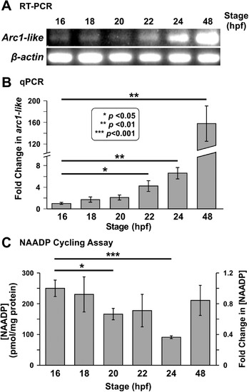
Expression of thearc1-liketranscript and detection of NAADP in whole zebrafish embryos from ~ 16 hpf to ~ 48 hpf. (A) RT-PCR followed by gel electrophoresis was conducted to show the relative expression of arc1-like in zebrafish embryos at ~ 16 hpf, ~ 18 hpf, ~ 20 hpf, ~ 22 hpf, ~ 24 hpf, and ~ 48 hpf (n = 4 for each time point). β-actin was used as an internal control. (B) Bar graph to show the mean ± SEM fold-change in arc1-likeexpression detected by quantitative real-time PCR (qPCR) relative to 16 hpf (n = 6). The expression of arc1-like was normalized to three house-keeping genes, actb2, ef1α, and rpl13a. (C) The NAADP cycling assay was employed to detect the endogenous level of NAADP in whole embryo extracts prepared at ~ 16 hpf, ~ 18 hpf, ~ 20 hpf, ~ 22 hpf, ~ 24 hpf, and ~ 48 hpf. This bar graph shows the normalized [NAADP] detected in the extract samples, and the corresponding fold-change in [NAADP] (n = 9). In (B) and (C), the asterisks indicate statistically significant differences at p < 0.05 (*), p < 0.01 (**) and p < 0.001 (***). In (C), the data obtained at ~ 18 hpf to ~ 48 hpf were compared statistically, with those obtained at ~ 16 hpf. |
| Gene: | |
|---|---|
| Fish: | |
| Anatomical Term: | |
| Stage Range: | 14-19 somites to Long-pec |
Reprinted from Developmental Biology, 445(2), Kelu, J.J., Webb, S.E., Galione, A., Miller, A.L., Characterization of ADP-ribosyl cyclase 1-like (ARC1-like) activity and NAADP signaling during slow muscle cell development in zebrafish embryos, 211-225, Copyright (2018) with permission from Elsevier. Full text @ Dev. Biol.