
Fig. 6
- ID
- ZDB-FIG-130128-17
- Publication
- Campbell et al., 2012 - Two types of tet-on transgenic lines for doxycycline-inducible gene expression in zebrafish rod photoreceptors and a gateway-based tet-on toolkit
- Other Figures
- All Figure Page
- Back to All Figure Page
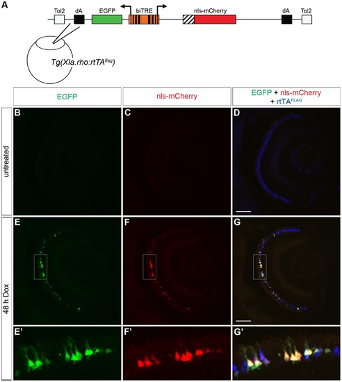
Bidirectional transactivation of an injected biTRE-containing plasmid into Tg(Xla.rho:rtTAflag). (A) Diagram of the bidirectional tetracycline response element (biTRE)-containing construct injected into Tg(Xla.rho:rtTAflag) one-cell embryos. EGFP and mCherry with a nuclear localization sequence (nls-mCherry) flank the biTRE. (B–G) Confocal z-projections of retinal sections from injected Tg(Xla.rho:rtTAflag) larvae at 6 dpf labeled with anti-FLAG antibody (blue). (B–D) GFP fluorescence (B, green) and nls-mCherry fluorescence (C, red) are undetectable in the absence of doxycycline (Dox) treatment, while anti-FLAG labeling (D, blue) is visible in rod photoreceptors. (E–G) GFP fluorescence (E, E2, green) and nls-mCherry fluorescence (F, F2, red) are visible in the photoreceptor layer and co-localize with anti-FLAG labeling (G, G2, blue) in the rod photoreceptors after 48 h Dox treatment. Boxed regions in E, F, and G correspond to E2, F2, and G2. dA, polyadenylation sequence; Tol2, pTol integration site. Scale bar (G), 50 µm. |