
Fig. 4
- ID
- ZDB-FIG-111128-46
- Publication
- Almeida et al., 2011 - Individual axons regulate the myelinating potential of single oligodendrocytes in vivo
- Other Figures
- All Figure Page
- Back to All Figure Page
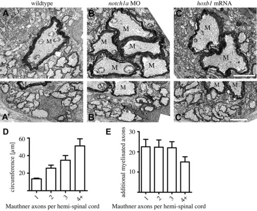
Supernumerary Mauthner axons are robustly myelinated. (A-C′) Transmission electron microscope images of transverse sections through the spinal cord of wild-type (A,A′), notch1a morphant (B,B′) and hoxb1 mRNA-injected (C,C′) zebrafish larvae at 9 dpf shows that all Mauthner axons are robustly myelinated (A-C) and that there is a similar number of axons myelinated in the ventral spinal cord despite the presence of supernumerary Mauthner (M) axons (A′-C′). Scale bars: 2 μm. (D) Total circumference of Mauthner axon(s) as a function of the number of Mauthner axons present per hemi-spinal cord. (E) Number of myelinated axons, excluding Mauthner axon(s), present in the ventral spinal cord as a function of the number of Mauthner axons present per hemi-spinal cord. Error bars represent s.d. |