
Fig. 1
- ID
- ZDB-FIG-110622-20
- Publication
- Cederlund et al., 2011 - mab21l2 transgenics reveal novel expression patterns of mab21l1 and mab21l2, and conserved promoter regulation without sequence conservation
- Other Figures
- All Figure Page
- Back to All Figure Page
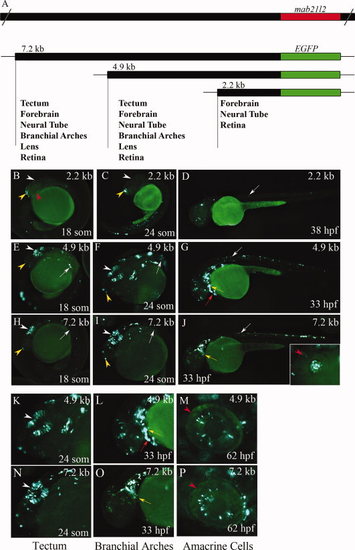
Identification of gene regulatory regions in zebrafish mab21l2. Schematic of the zebrafish mab21l2 gene, and the mab21l2 promoter-reporter constructs and their expression domains (A). Epifluorescent images of wholemount larvae injected with mab21l2:EGFP promoter constructs (B–P). The 2.2-kb construct shows some activity in the forebrain and neural tube (B–D). In contrast, the 4.9- and 7.2-kb constructs direct expression in most known mab21l2expression domains (E–P). At <18–24 somites, they direct EGFP expression in the developing optic tectum, forebrain, and neural tube (E,F,H,I,K,N). At <25 hpf, both promoter constructs drive expression in the lens (inset in J). At <33 hpf, EGFP is still expressed by cells in the optic tectum, branchial arches, neural tube, and in tissue surrounding the heart (G,J,L,O). At <62 hpf, EGFP-positive cells are apparent in the eye (M,P). White arrowheads, tectum; white arrows, neural tube; red arrowheads, eye; red arrows, heart; yellow arrowheads, forebrain; yellow arrows, branchial arches. F,I,K,N, dorsal views; B–D,E,G,H,J,L,M,O,P, lateral views. |