
Fig. 1
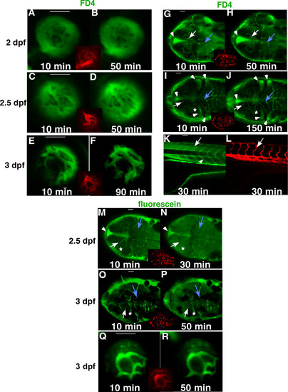
Development of the BRB and BBB in zebrafish. FD4 (4,000 Da, green) was injected to the sinus venosus of Tg(flk1:mCherry) embryos at 2 dpf (A, B, G, H), 2.5 dpf (C, D, I, J) and 3 dpf (E, F, K. L). Live confocal images of the hyaloid vessels (A-F, side view), brain vessels (G-J, dorsal view) and trunk vessels (K-L, side view) were obtained from 10 to 150 minutes after injection. Insets and panel L are images of established vasculature of the injected embryos (red). In the brain, boundaries of the middle mesencephalic central artery (MMCtA) are indicated by arrows in G-J, cerebellar central artery (CCtA), by blue arrows in G-J and posterior mesencephalic central artery (PMCtA) by asterisks in I&J. Arrows in K&L indicate intersegmental vessels and arrowhead in K shows the myotomal boundaries. A small molecule, fluorescein (376 Da, green), was also used as a tracer in the injection assay (M-R). In the 2.5 dpf embryos (M&N), the PMCtA (asterisk) could not be differentiated, and boundaries of MMCtA (arrows) and CCtA (blue arrows) were enlarged and became blurred 30 minutes after injection. However in the 3 dpf embryos injected with fluorescein (O-R), boundaries of the MMCtA (arrows), PMCtA (asterisk), CCtA (blue arrows) and the hyaloid vessels (Q&R) remained sharp and clear after 50 minutes of injection. The leaked FD4 accumulated mostly in brain ventricles (arrowheads in G-J). In contrast, most of the leaked fluorescein did not accumulated in the brain ventricle (arrowheads in M&N), but evenly diffused throughout the brain (M-P). Scale bars: 50 μm. |