
- Title
-
Genetic and protein interaction studies between the ciliary dyslexia candidate genes DYX1C1 and DCDC2
- Authors
- Bieder, A., Chandrasekar, G., Wason, A., Erkelenz, S., Gopalakrishnan, J., Kere, J., Tapia-Páez, I.
- Source
- Full text @ BMC Mol Cell Biol
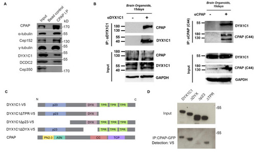
CPAP interacts with DCDC2 and DYX1C1. (A) CPAP endogenously interacts with DCDC2 and DYX1C1. Endogenous immunoprecipitations using HeLa cell extracts. CPAP was used as a bait to pull down interactors. CEP350 was used as a negative control and CEP152 was used as a positive control for interaction with CPAP. Beads alone were used as a negative control for the IP´s. (B) CPAP and DYX1C1 interaction in brain organoids. CPAP was immunoprecipitated by anti- DYX1C1 in 15 day old brain organoids. Reciprocally, DYX1C1 was pulled down by anti-CPAP. (C) Schematic representation of domain structures of DYX1C1, CPAP and the deletion constructs DYX1C1ΔTPR, DYX1C1Δp23, DYX1C1ΔDYX. p23 = p23 domain, TPR = tetratricopeptide repeat domain, DYX = DYX domain. (D) CPAP interacts with DYX1C1 via the p23 domain. hTERT-RPE1 cells stably expressing DOX-CPAP-GFP and growing in normal serum conditions were induced with doxycycline and transiently transfected with the indicated constructs. GFP-Trap was used to pull down CPAP-GFP. anti-V5 antibody was used for immunodetection of interactors. |

Co-occurrence of DYX1C1 and CPAP at the subcellular level. A) DYX1C1 and CPAP co-occur around the centrosome. Confocal immunofluorescence images of endogenous protein in hTERT-RPE1 cells grown in normal serum conditions. B)-C) DCDC2 co-occurs with CPAP depending on cell cycle stage. Nuclei are stained with DRAQ5. Scale bar = 10 μm |
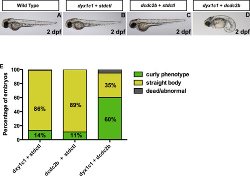
Genetic interaction between dyx1c1 and dcdc2b in zebrafish. A,B,C) Injection of 20 µM of dyx1c1MO or 100 µM of dcdc2bMO individually in zebrafish embryos resulted in a maximum number of embryos with straight body axis similar to wildtype embryos at 2dpf. D) Co-injection of the subphenotypic doses of dyx1c1MO and dcdc2bMO (20 µM dyx1c1 + 100µM dcdc2b) resulted in a strong curved body axis in a greater number of embryos at 2dpf. StdctlMO was used in order to maintain a uniform concentration of the morpholinos. E) Graphical representation of percentage of embryos with strong curly phenotype following MO injection. Coinjection of dyx1c1 and dcdc2b resulted in a dramatic increase in number of embryos with strong curly body axis as compared to individual knockdown of the two genes. The number of embryos examined in control and knockdown groups were 100 (n = 100). PHENOTYPE:
|

Transcriptional control between DYX1C1/dyx1c1 and DCDC2/dcdc2b. |