
|
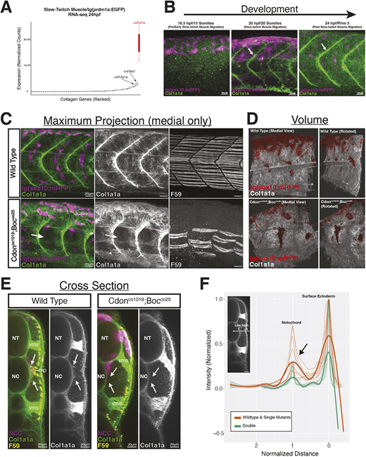
Fig. 7 Col1a1a matrix deposition in cdon;boc mutants. (A) RNA-seq from FAC sorted tg(prdm1a:eGFP) cells at 24 hpf identify expression of collagen genes. (B) 3D projections of Col1a1a deposition at the level of yolk extension. At 20 hpf, Col1a1a expression is strongest in the vertical myosepta (arrows). (C) Medial view shows Col1a1a deposition along the somite-notochord boundary in wild-type and double mutant embryos at 24 hpf. Col1a1a signal in skin ectoderm was removed for images in C. The loss of Col1a1a in the double mutant is associated with loss of slow-twitch muscle differentiation (arrows). Retention of Col1a1a around dorsal neural tube (arrows) coincides with the location of tNCC migration arrest. (D) Volume rendering shows medial deposition of Col1a1a in wild-type (top) and double mutant (bottom) embryos at 24 hpf. (E) Optical cross-section shows loss of medial Col1a1a deposition along the NCC migratory path (arrows) in double mutant embryos at 24 hpf. (F) Line scan quantification showing reduced Col1a1a intensity in double mutant embryos. Line scans taken on optically sectioned images and normalized for length and intensity. NC, notochord; NT, neural tube; mp, muscle pioneers; vms, vertical myosepta.