
|
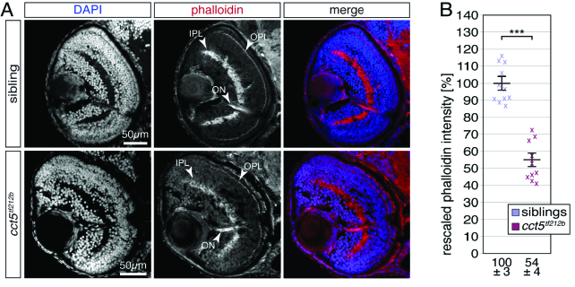
Figure 3 F-actin is significantly reduced in the retina of cct5𝑡𝑓121𝑏 mutants. (A) At three dpf, F-actin was labelled with phalloidin (red) on cross sections counterstained with DAPI (blue). Whereas the inner and outer plexiform layers of the retina (IPL and OPL, respectively) were prominently stained by phalloidin within siblings, the signal from cct5𝑡𝑓121𝑏 homozygotes appeared weaker. Phalloidin also marked the optic nerve (ON). (B) Quantification of the brightness of the phalloidin signal within the IPL revealed that cct5𝑡𝑓121𝑏 homozygotes showed a significant reduction in signal intensity to 54 ± 4% in relation to their siblings, which were rescaled to 100 ± 3%. Data are mean ± SEM; ***P < 0.001 by Student’s t-test; n = 10.