
|
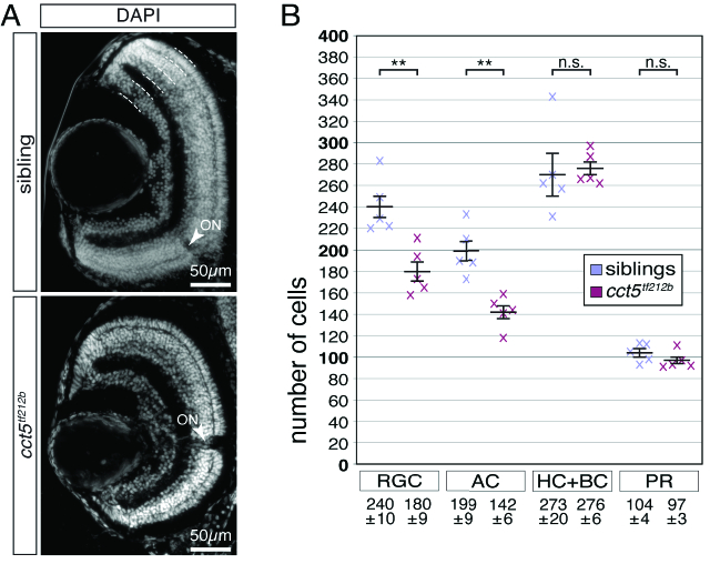
Figure 4 Retinal ganglion and amacrine cells are reduced in number in cct5𝑡𝑓121𝑏 mutants. (A) At six dpf, the retinal nuclei of siblings and cct5𝑡𝑓121𝑏 mutants were marked at the level of the optic nerve using the nuclear stain DAPI. Indicated by the dotted lines, the outer nuclear layer contains the photoreceptor cells (PR). The inner nuclear layer harbours the apical horizontal cells (HC) and the bipolar cells (BC) as well as the basally located amacrine cells (AC). The most basal layer contains the retinal ganglion cells (RGC). Arrowheads indicate optic nerves (ON). (B) Quantification of the cellularity of the different retinal layers within siblings and cct5𝑡𝑓121𝑏 homozygotes at six dpf. The cell number of the RGC and the AC were significantly reduced in cct5𝑡𝑓121𝑏 homozygotes. Per retinal cross section, siblings had 240 ± 10 RGC and 199 ± 9 AC in contrast to 180 ± 9 RGC and 142 ± 6 AC in cct5𝑡𝑓121𝑏 homozygotes. The number of HC and BC as well as PR remained unchanged. Siblings had 273 ± 20 HC and BC as well as 104 ± 24 PR and cct5𝑡𝑓121𝑏 homozygotes had 276 ± 6 HC and BC as well as 97 ± 3 PR. Data are mean ± SEM; **P < 0.01 by Student’s t-test; n.s., not significant; n, 5.