
Image
|
Figure Caption
Fig. 4
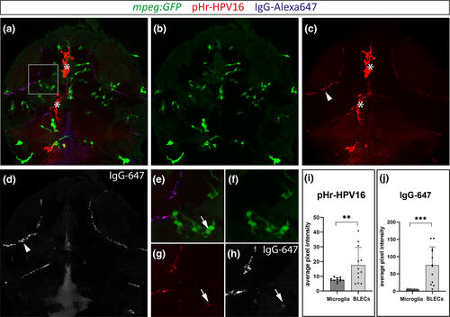
BLECs ingest HPV-16 pseudoviruses more efficiently than microglia. (a-d) Maximum projection of a mpeg:GFP positive zebrafish embryo that was co-injected with pHr-HPV-16 and IgG-Alexa647 into the CSF at 5dpf. The white box indicates the zoom-in area in (e-h). Single channel of maximum projection in (a) for mpeg:GFP (b), pHr-HPV-16 (c) and IgG-Alexa647 (d). Arrowheads highlight uptake into BLECs while arrows mark endocytosis of the respective substrate in microglia. Note the auto-fluorescence of pigment cells in the red channel, marked by asterisks. (i, j) Quantification of the average pixel intensities within mpeg:GFP+ microglia and BLECs in n = 12 embryos shows a significantly higher uptake of pHr-HPV-16 (Welch's t-test, p = .0097) and IgG-Alexa647 (Welch's t-test, p = .0001) in BLECs when compared to microglia. Values are presented as means ± SD
Figure Data
Acknowledgments
This image is the copyrighted work of the attributed author or publisher, and
ZFIN has permission only to display this image to its users.
Additional permissions should be obtained from the applicable author or publisher of the image.
Full text @ Glia