IMAGE

Fig. 2

ID
ZDB-IMAGE-220727-2
Genes
Source
Figures for Wang et al., 2022
Image
Figure Caption

Fig. 2

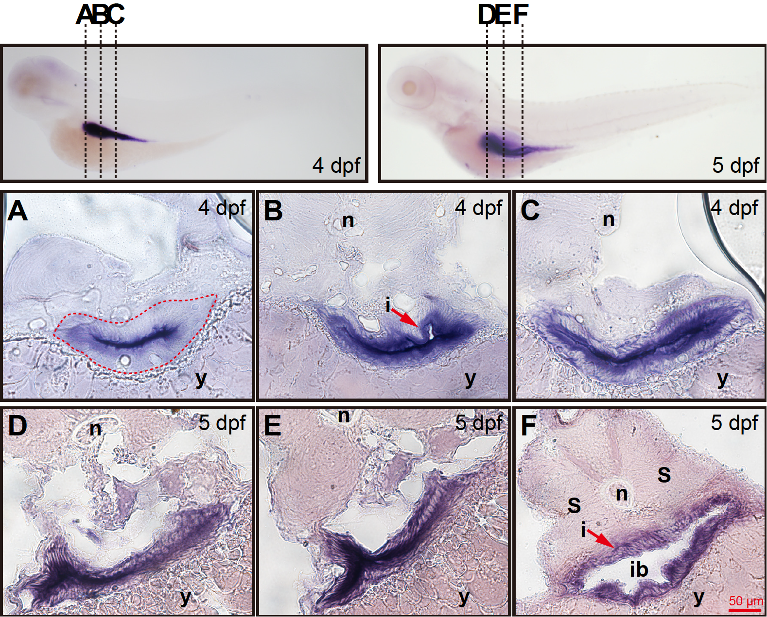
(A–C) Corresponding transverse sections through the three different regions of intestine at 4 dpf depicted in left image of the top row. (A) The aqp8ab mRNA was mainly expressed in epithelial cells lining the intestinal cavity. The developing intestine adjacent to the yolk was seen at 4 dpf (red dotted lines). (B) The aqp8ab mRNA expression was more intense in the layer lining the intestinal cavity. The intestinal tract contained a small lumen (arrow). (C) The aqp8ab mRNA expression was still detected in the layer lining the intestinal cavity. (D–F) Corresponding histological cross-sections through the different regions of intestine at 5 dpf depicted in right image of the top row. Cross-sections analysis showed that aqp8ab mRNA expression was more intense in the layer lining the intestinal lumen. n, notochord; i, intestine; s, somite; y, yolk; ib, intestinal blub.

Figure Data
Acknowledgments
This image is the copyrighted work of the attributed author or publisher, and ZFIN has permission only to display this image to its users. Additional permissions should be obtained from the applicable author or publisher of the image. Full text @ Acta. Biochim. Biophys. Sin (Shanghai)