IMAGE

Fig 4

ID
ZDB-IMAGE-210817-14
Source
Figures for Bengani et al., 2021
Image
Figure Caption

Fig 4 Expression levels of <italic toggle='yes'>Fmr1</italic> and FMRP in <italic toggle='yes'>Fmr1</italic><sup>CRE</sup>.

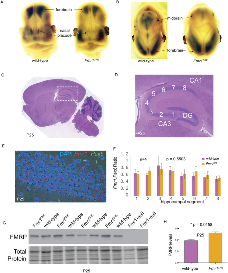
Frontal (A) and saggital (B) views of 13.5GD embryonic mouse heads following whole-mount in situ hybridization for Fmr1. In each panel the wild-type male embryo is shown on the left and the Fmr1CRE embryo on the right. There is loss of expression of Fmr1 in the nasal placode and midbrain Fmr1CRE mutant embryos as compared to wild-type embryos. The Fmr1CRE embryos had been deliberately over-developed in the chromogenic substrate compared to the wild-type embryos to emphasize the signal difference. Saggital H&E stained section of whole brain (C) with detailed view (white dashed box) of the hippocampus (D) with marked hippocampus regions indicating the regions analysed in (F) numbered 1–8, starting from dentate gyrus. (E) Reference image of RNAscope processed section with Fmr1 transcript (red), Pax6 transcript (green) and nucleus (blue/DAPI). Each transcript is represented by a spot following the quantitative image processing. (F) Graphical representation of Fmr1 transcripts normalised to Pax6 transcripts (used as control) between Fmr1CRE (purple) compared to wild-type littermates (orange) and data represent average of four replicates (n = 4) ±SE. Levels of significance were determined by 2-tailed Student’s t-test, with p values lower than 0.05 considered statistically significant. No significant difference was observed in the Fmr1 transcript levels. (G) Western blot of hippocampal tissue from four Fmr1CRE, four wild-type and two Fmr1-null mice at P25 using an antibody that detects FMRP. (H) Quantitation of the FMRP bands in (G) indicating an apparent increase in FMRP in Fmr1CRE hippocampal slices. All quantitative data are presented as mean ±SE and p value of 0.05 or less is considered statistically significant. (* means difference is statistically significant). FB: Forebrain; MB: Midbrain; NP: Nasal placode; DG: Dentate gyrus.

Acknowledgments
This image is the copyrighted work of the attributed author or publisher, and ZFIN has permission only to display this image to its users. Additional permissions should be obtained from the applicable author or publisher of the image. Full text @ PLoS One