IMAGE

Fig 2

ID
ZDB-IMAGE-210113-119
Source
Figures for Nunley et al., 2020
Image
Figure Caption

Fig 2 A Y-Junction, a topological defect in the cone mosaic, is an insertion of a row and a column.

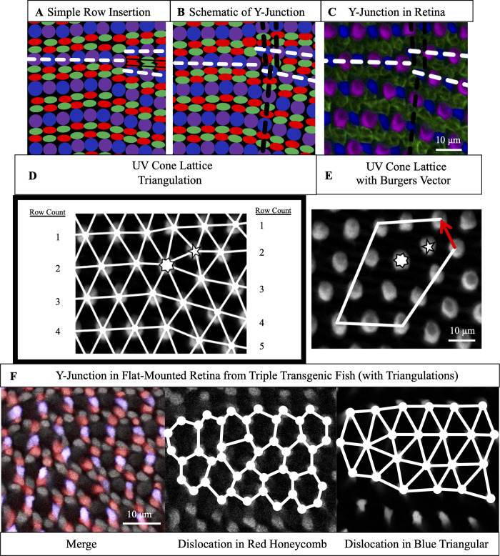
(A) Schematic of simple row insertion in cone mosaic. As new cone photoreceptors are incorporated to the right of the defect, a series of improper cone contacts (black box) within columns form. White dashed lines: rows associated with defect. (B) Schematic of a Y-Junction, a topological defect in the zebrafish cone mosaic. A Y-Junction only disrupts the cone mosaic near the core rather than along an entire line of contacts. White (black) dashed lines: rows (columns) associated with the defect. (C) A Y-Junction in a flat-mount retinal preparation from an adult, double transgenic (Tg[sws1:GFP; sws2:mCherry]) line in which UV and Blue cones express distinct fluorescent reporters (pseudo-colored magenta and blue, respectively) under control of UV and Blue opsin promoters, respectively. Antibody staining labels Red and Green cones (both pseudo-colored green). White (black) dashed lines: rows (columns) associated with the defect. (D) Each UV cone from panel C is connected (white bonds) to its nearest UV cone neighbors. To the left and right of the defect, rows are counted. Seven-sided (five-sided) star: seven-coordinated (five-coordinated) UV cone. (E) A circuit of triangulation bonds around defect from panels C-D. Red arrow is the Burgers vector, the additional bond necessary to close the circuit containing a dislocation. (F) Y-Junction in the same flat-mount retinal preparation as in Fig 1B. The round cells with dim fluorescence are UV cones. The Red cones are pseudo-colored red. The Blue cones are pseudo-colored blue. The remaining cones (bright gray fluorescence) are Green cones. We connect (solid white lines) nearest neighbors in the Red cone sublattice and Blue cone sublattice.

Acknowledgments
This image is the copyrighted work of the attributed author or publisher, and ZFIN has permission only to display this image to its users. Additional permissions should be obtained from the applicable author or publisher of the image. Full text @ PLoS Comput. Biol.