
Image
|
Figure Caption
Figure 6
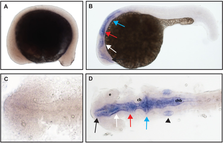
(A and C) At 16.5 hpf, no expression was detected. (B and D) Expression was seen in the telencephalon (black arrows), the diencephalon (white arrows), the midbrain (red arrows), the hindbrain (blue arrows), and otic vesicle (black arrowhead). (A and B) Lateral view. (C and D) Dorsal view. e, marks the eye, cb, marks the cerebellum, and chb, marks the caudal hindbrain. All embryos are oriented with the head to the left. This data is representative of the results of three rounds of in situ hybridization with at least 25 embryos per round.
Figure Data
Acknowledgments
This image is the copyrighted work of the attributed author or publisher, and
ZFIN has permission only to display this image to its users.
Additional permissions should be obtained from the applicable author or publisher of the image.
Full text @ Peer J.