
|
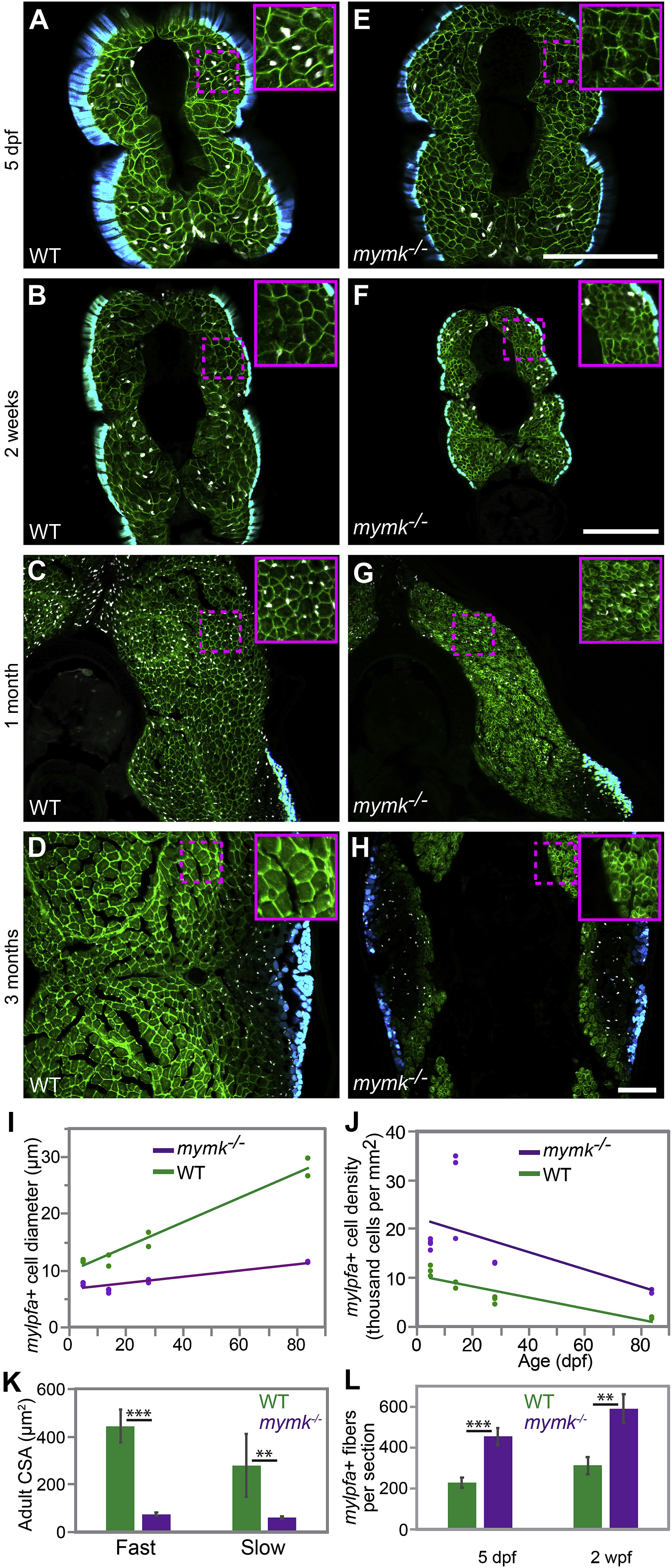
Fig. 4 mymk is required for normal myofiber growth. Transverse sections through the mid-trunk region of wild-type (WT) (A–D) and mymkoz17 (E–H) 3MuscleGlow transgenic fish at 5 days, 2 weeks, 1 month, and 3 months post-fertilization. myog:H2B-mRFP (white) labels myonuclei, mylfpa:lyn-cyan (green) marks fast muscle cell membranes, and smyhc1:EGFP (aqua) marks slow muscle cells. Insets show magnified images of regions indicated by the dotted box. (I) Diameter of mylpfa-expressing fast muscle cells in WT and mymkoz17 individuals at the same stages, with trends over time determined by linear regression (model R2 is 0.97). (J) Fast muscle cell density is shown for the same WT (green) and mymkoz17 (purple) individuals as in (I) and regression lines show trends through time (model R2 is 0.63). Linear regression was performed in JMP using standard least squares modeling. (K) Adult myofiber cross-sectional area is higher in WT than mymkoz17 in both slow and fast myofiber types. (L) Total myofiber counts per section are lower in WT than in mymkoz17 at 5 dpf and 2 wpf. Scale bars in E (for A, E), in F (for B, F, C, G), and in H (for D, H) are 100 μm. Student’s t-test, p∗∗<0.01, p∗∗∗< 0.001.
Reprinted from Developmental Biology, 462(1), Hromowyk, K.J., Talbot, J.C., Martin, B.L., Janssen, P.M.L., Amacher, S.L., Cell fusion is differentially regulated in zebrafish post-embryonic slow and fast muscle, 85-100, Copyright (2020) with permission from Elsevier. Full text @ Dev. Biol.