
|
Fig. 4
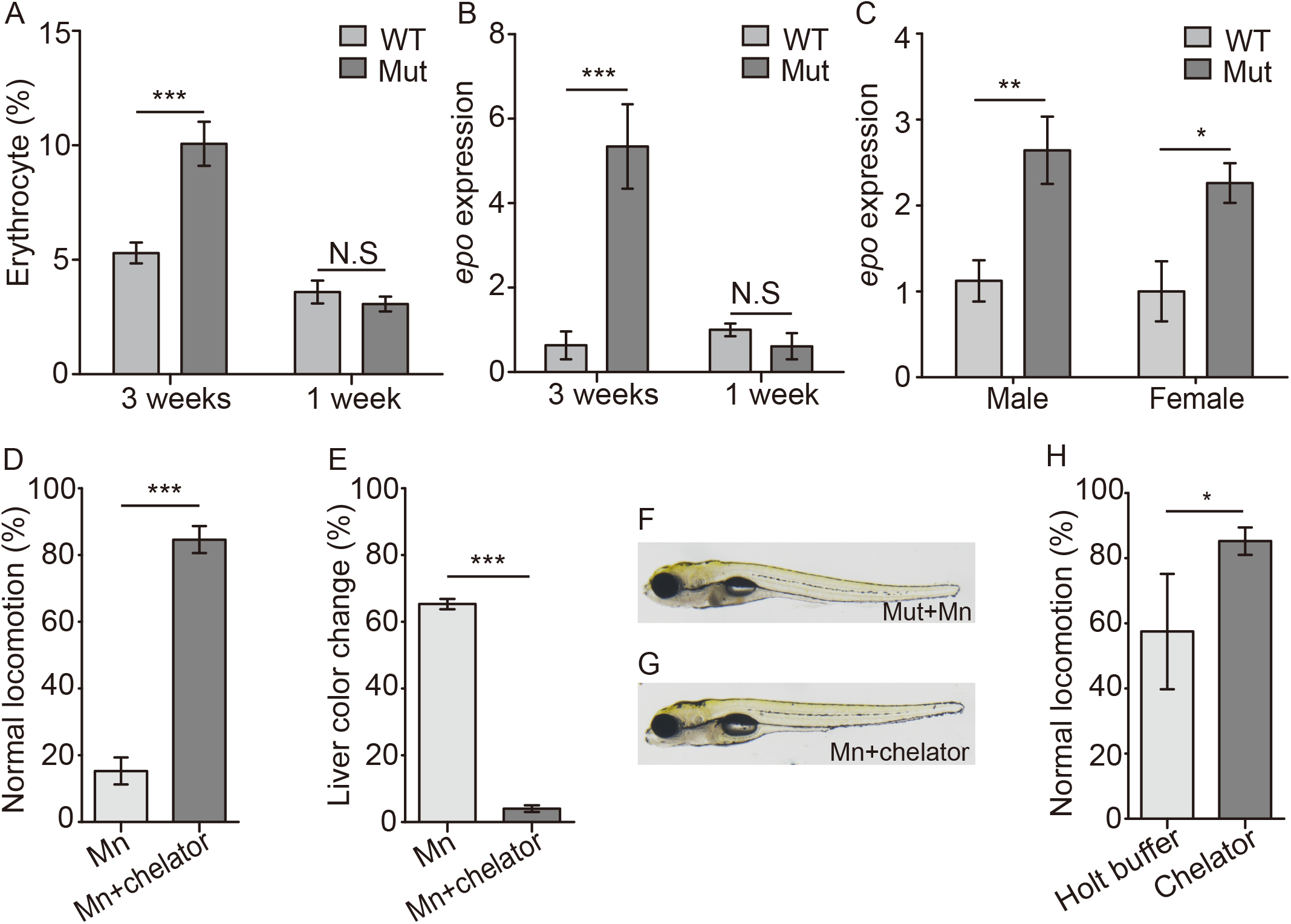
Slc30a10 mutant zebrafish develop polycythemia, and chelation treatments improves the phenotype in mutant embryos.
(A) Summary of the relative numbers of erythrocytes in 3-week-old and 1-week-old embryos (n = 6 sets of 50 embryos/group). (B and C) Epo expression was measured in 3-week-old and 1-week-old mutant zebrafish (B) and in the liver of adult zebrafish (C); n = 3 sets of 20 adults/group. (D and E) Mn-induced locomotor defects (D) and the darker color in liver (E) were rescued by treating embryos with EDTA-CaNa2 (chelator, n = 3 sets of 20 embryos/group). (F-G) Example images of a Mn-exposed mutant embryo, showing a darker colored liver (F), and a Mn-exposed mutant embryo following EDTA-CaNa2 treatment (G). (H) Following exposure to 300 μM Mn, mutant embryos were transferred to either Holt buffer alone or Holt buffer containing EDTA-CaNa2, and locomotion was measured (n = 3 sets of 20 embryos/group). *p<0.05, **p<0.01, and ***p<0.001.