
|
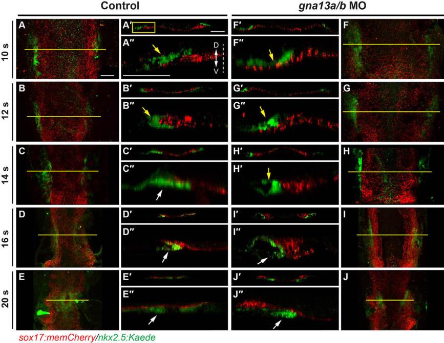
Fig. S3
Gα13 is required for subduction and the final stage of the medial migration of myocardial precursors. (A-J) Projections of confocal Z-stacks of anterior endoderm and myocardial precursors (taken by a Lan-Apo 20×/NA 0.8 objective) in embryos at different stages as indicated, showing the mCherry-labeled endoderm (red) and Kaede-labeled myocardial cells (green). (A'-J') Images of XZ transverse sections in the areas, as indicated by yellow lines in A-J. (A''-J'') Magnified areas of the left region of the XZ transverse sections in A'-J'. Example of region used is indicated by box in A'. Dashed line: midline; arrows: myocardial cells, located above (yellow) and below (white) the endoderm; D: dorsal; V: ventral. Scale bars: 20 μm.