
Image
|
Figure Caption
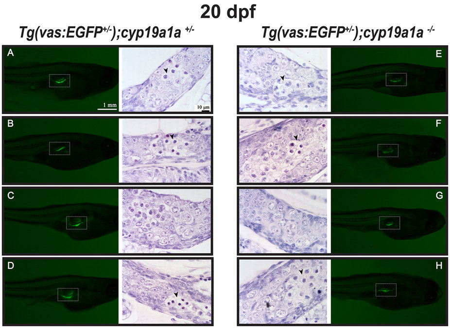
Fig. 4
Gonad development at 20 dpf in the control [Tg(vas:EGFP);cyp19a1a+/−; fish A–D] and mutant [Tg(vas:EGFP);cyp19a1a−/−; fish E–H]. Similar GFP signals (boxed in the photo) were observed in the two groups and histological examination showed no significant difference. Meiotic germ cells (arrowhead) with condensed chromatin were often seen in both the mutant and control gonads.
Figure Data
Acknowledgments
This image is the copyrighted work of the attributed author or publisher, and
ZFIN has permission only to display this image to its users.
Additional permissions should be obtained from the applicable author or publisher of the image.
Full text @ Sci. Rep.