
Image
|
Figure Caption
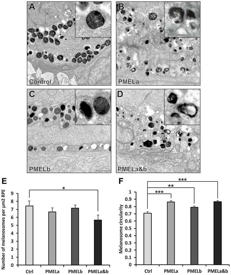
Fig. 5
PMELa MO reduces the number of cylindrical melanosomes to a greater extent than PMELb MO. (A–D) Electron micrographs highlighting melanosome morphology at 2dpf for zebrafish injected with control (A), PMELa (B), PMELb (C) and PMELa&b MOs (D). Scale bars: 1µm. (E) The PMELa&b MO resulted in a decrease in the number of melanosomes. Ctrl, control. (F) All PMEL MOs resulted in a significant loss of cylindrical melanosomes. PMELa had a greater effect on melanosome shape than the PMELb MO. Results show the mean±s.e.m. *P<0.05, **P<0.01, ***P<0.001 (Student′s t-test).
Figure Data
Acknowledgments
This image is the copyrighted work of the attributed author or publisher, and
ZFIN has permission only to display this image to its users.
Additional permissions should be obtained from the applicable author or publisher of the image.
Full text @ J. Cell Sci.