
|
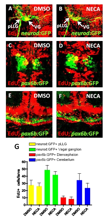
Fig. S4
NECA does not increase proliferation of neuronal cells during β cell regeneration, related to Figure 4.
(A-B) Proliferation of cells in neuronal ganglia was assessed by EdU incorporation in Tg(neurod:GFP)-expressing cells in the posterior lateral line ganglion (pLLG) and vagal ganglion (VG). Tg(neurod:GFP);Tg(ins:flag-NTR) larvae were treated with MTZ from 3-4 dpf for ablation of the β cells, and subsequently treated with DMSO (A) or NECA (B) and EdU during β cell regeneration from 4-6 dpf.
(C-F) Proliferation of neuronal cells in the central nervous system was assessed by EdU incorporation in Tg(pax6b:GFP)-expressing cells in the diencephalon (C-D) and cerebellum (E-F). Tg(pax6b:GFP);Tg(ins:flag-NTR) larvae were treated with MTZ from 3-4 dpf for ablation of the β cells, and subsequently treated with DMSO (E) or NECA (F) and EdU during β cell regeneration from 4-6 dpf. Confocal projections are shown in (A-F) to clearly visualize the structures although EdU incorporation was assessed on each individual plane.
(G) Quantification of the number of cells that incorporated EdU in each neuronal compartment. No significant difference was found between DMSO- and NECA-treated larvae. n = 8-13 larvae for each group. Error bars represent SEM.
Reprinted from Cell Metabolism, 15(6), Andersson, O., Adams, B.A., Yoo, D., Ellis, G.C., Gut, P., Anderson, R.M., German, M.S., and Stainier, D.Y., Adenosine Signaling Promotes Regeneration of Pancreatic beta Cells In Vivo, 885-894, Copyright (2012) with permission from Elsevier. Full text @ Cell Metab.