
|
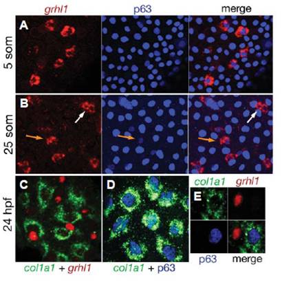
Fig. 2 grhl1 is transiently expressed in a subset of epidermal precursors, but absent from differentiated basal keratinocytes. (A,B)
Fluorescent in situ hybridisation for grhl1 (in red) combined with immunostaining for p63 (in blue). (C) Double fluorescent in situ hybridization for col1a1 (in green) andgrhl1 (in red). (D) Fluorescent in situ hybridisation for col1a1 (in green) combined with immunostaining for p63 (in blue). (E) A triple staining for col1a1 mRNA, grhl1 mRNA and p63 protein (three single channels plus merged image). For panels (C-E), embryos injected with grhl1 splice MO were used, leading to accumulation of grhl1 mRNA in the nucleus and facilitating the analysis of the obtained patterns (compare with Figs. 6 and S7).