
Image
|
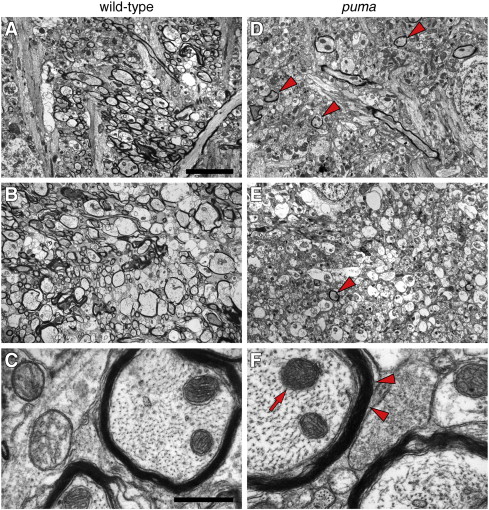
Figure Caption
Fig. 7 Reduced myelination in puma mutant brains revealed by transmission electron microscopy. (A–C) Representative regions of wild-type at low magnification (5000×, A and B) and higher magnification (60,000×, C) show numerous well-myelinated axon tracts. (D–F) Corresponding regions and magnifications in puma reveal far fewer myelinated axon tracts (arrowheads in D and E) though layering of individual myelin sheaths that are present is indistinguishable from wild-type (arrowheads in F; arrow, mitochondrion). Scale bars: in A, 5 μm, for A, B, D, E; in C, 500 nm for C, F.
Figure Data
Acknowledgments
This image is the copyrighted work of the attributed author or publisher, and
ZFIN has permission only to display this image to its users.
Additional permissions should be obtained from the applicable author or publisher of the image.
Reprinted from Developmental Biology, 346(2), Larson, T.A., Gordon, T.N., Lau, H.E., and Parichy, D.M., Defective adult oligodendrocyte and Schwann cell development, pigment pattern, and craniofacial morphology in puma mutant zebrafish having an alpha tubulin mutation, 296-309, Copyright (2010) with permission from Elsevier. Full text @ Dev. Biol.