
Image
|
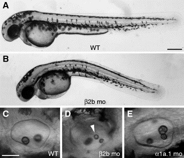
Figure Caption
Fig. 5 Knockdown of Na,K-ATPase β2b expression. All panels show a lateral view with anterior to the left. (A) Wild type (WT) embryo at 45 hpf. (B) Embryo injected with 6 ng of β2b MO-1 at 45 hpf. (C) Otic vesicle (OV) of WT embryo at 45 hpf. (D) OV of embryo injected with 6 ng of β2b MO-1 at 45 hpf. Arrowhead indicates ectopic otolith. (E) OV of embryo injected with 0.125 ng of α1a.1 MO-1 at 45 hpf. mo, morphant. Scale bars: A–B, 250 μm; C–H, 50 μm.
Acknowledgments
This image is the copyrighted work of the attributed author or publisher, and
ZFIN has permission only to display this image to its users.
Additional permissions should be obtained from the applicable author or publisher of the image.
Reprinted from Developmental Biology, 294(1), Blasiole, B., Canfield, V.A., Vollrath, M.A., Huss, D., Mohideen, M.A., Dickman, J.D., Cheng, K.C., Fekete, D.M., and Levenson, R., Separate Na,K-ATPase genes are required for otolith formation and semicircular canal development in zebrafish, 148-160, Copyright (2006) with permission from Elsevier. Full text @ Dev. Biol.